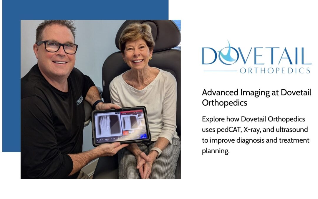
When it comes to orthopedic care, pinpointing the exact cause of pain or dysfunction isn’t always straightforward. That’s because muscles, tendons, joints, and bones are all deeply connected—meaning multiple issues can exist at once or mask one another. A basic image might show a fracture, but not the misalignment. A joint might look fine in one position, but fail under stress. This is why Dovetail Orthopedics has invested in advanced imaging tools that go beyond the surface. Our combination of pedCAT™ 3D CT scans, traditional X-rays, and diagnostic ultrasound allows us to deliver faster, more accurate diagnoses—and better outcomes.
pedCAT CT Scan: 3D Precision for Foot and Ankle
pedCAT™ is a specialized CT scanner designed specifically for the foot and ankle. Unlike standard X-rays, which offer only two-dimensional views, pedCAT creates ultra-thin cross-sectional slices that can be digitally reassembled into a full 3D image. This level of detail allows our team to examine every angle of the bones and joints in your foot and ankle, uncovering hidden issues that might be missed otherwise.
We use pedCAT to diagnose and evaluate:
- Fractures of the foot and ankle
- Arthritis and joint degeneration
- Pre-operative surgical planning
- Post-surgical bone healing
- Dislocations and misalignments
- Non-unions and delayed healing
- Angular deformities and poor joint relationships
Whether you’re recovering from an injury or planning a corrective procedure, pedCAT gives us the most complete picture possible—literally.
X-Ray Imaging: A Diagnostic Staple
X-rays remain one of the most effective and immediate tools for diagnosing orthopedic conditions. At Dovetail, we use digital X-rays to evaluate bone fractures, joint spaces, and structural alignment. These quick and painless images are often the first step in determining whether more advanced imaging is needed.
Our X-rays help assess:
- Acute fractures
- Bone spurs and degenerative changes
- Joint alignment
- Post-operative hardware placement
- Foreign objects or calcifications
While they don’t show soft tissues like tendons or ligaments, X-rays provide critical baseline information for diagnosing and tracking bone-related conditions.
Ultrasound Imaging: Real-Time Insight into Soft Tissues
Muscle strains, tendon tears, cysts, and inflammation don’t always show up clearly on X-rays or CT scans. That’s where ultrasound comes in. At Dovetail Orthopedics, we use high-resolution ultrasound to evaluate soft tissue in real time—watching how tendons glide, identifying fluid buildup, or locating the precise point of pain.
Ultrasound offers:
- Dynamic, real-time visualization
- No radiation exposure
- Immediate feedback for guided injections
- In-office convenience for quicker answers
It’s especially useful for diagnosing plantar fasciitis, tendonitis, neuromas, and other non-bony injuries. When combined with our other imaging tools, ultrasound helps round out the full picture of your orthopedic health.
Get the Right Diagnosis, the First Time
If you’ve been living with pain or uncertainty, don’t settle for guesswork. At Dovetail Orthopedics, our advanced imaging technology—pedCAT, X-rays, and ultrasound—gives us the clarity to diagnose accurately and build a treatment plan that works.Take a minute to meet the team and learn more about Dovetail Orthopedics.