Dovetail Orthopedics now offers state of the art
Standing
CT Imaging
Your doctor may determine your X-Ray does not provide enough information about your injury and may order a standing CT scan. CT scans provide a complete 3D picture of your bone alignment and joint spaces.
The American Orthopedic Foot & Ankle Society recommends standing (weight bearing) imaging, when possible, to get the most accurate assessment of the functional bony anatomy of the foot and ankle.¹
Deformities of the forefoot, midfoot, and hindfoot have been shown to be more visible in standing position.²

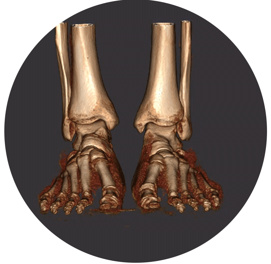
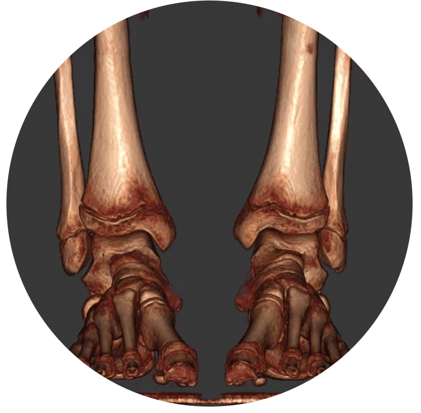
The standing, weight-bearing CT of the ankle (left) shows where bones are making contact when the patient is standing. This information guides the surgeon on the extent of the patient’s deformity and is useful in determining the best treatment plan. Traditional supine, non-weight bearing CT (right) may not show the deformity since the patient is lying down.
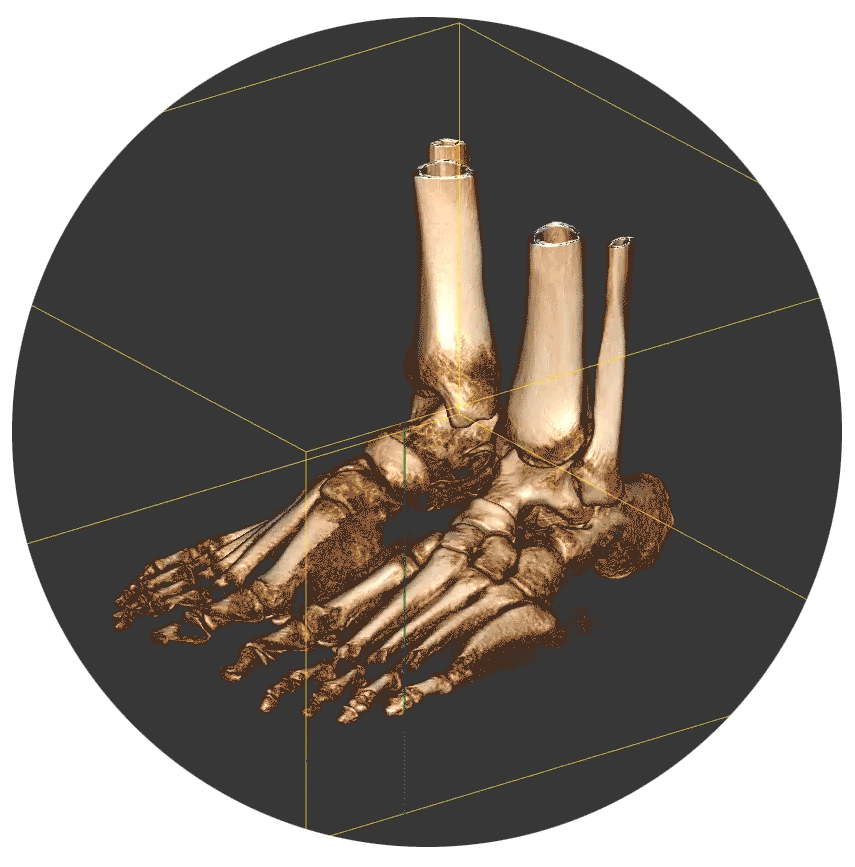
Three-dimensional imaging helps ensure the right diagnosis the first time.


Standing CT is perfect for:
- Pre-operative planning
- Post-operative assessment
- Diagnosis of fractures
- Evaluation of arthritic joints
- Evaluation of bunion deformity
- Evaluation of ankle instability
- Evaluation of foot alignment
- Sesamoid position and condition
Why
standing CT?
- Diagnose fractures with more accuracy³
- Evaluate joint wear (osteoarthritis) and
deformities of the forefoot (hallux
valgus, claws, hammer toes), midfoot
and hindfoot - Create precise surgical plans⁴
- Evaluate post-operative healing

Standing CT images provide:
3 Dimensional views
Weightbearing Diagnosis
Low Radiation Exposure
FAQ


Standing CT combines the benefits of X-Ray and Full-Body CT.
Weightbearing CT is the Clear Choice
¹Choosing Wisely: Five Things Physicians and Patients Should Question”. American Orthopaedic Foot & Ankle Society. Released September 17, 2014. For more information, visit www.choosingwisely.com.
²Conti MS, Ellis SJ. Weight-bearing CT Scans in Foot and Ankle Surgery. J Am Acad Orthop Surg. 2020 Jul 15;28(14):e595-e603. doi: 10.5435/JAAOS-D-19-00700. PMID: 32692095.
³Ricci PM, Boldini M, Bonfante E, Sambugaro E, Vecchini E, Schenal G, Magnan B, Montemezzi S. Cone-beam computed tomography compared to X-ray in diagnosis of extremities bone fractures: A study of 198 cases. Eur J Radiol Open. 2019 Mar 13;6:119-121. doi: 10.1016/j.ejro.2019.01.009. Erratum in: Eur J Radiol Open. 2020 Dec 17;8:100308. doi: 10.1016/j.ejro.2020.100308. PMID: 30911591; PMCID: PMC6416521.
⁴Chun, D.-I., Cho, J., Won, S. H., Nomkhondorj, O., Kim, J., An, C. Y., & Yi, Y. (2025). Weight-Bearing CT: Advancing the Diagnosis and Treatment of Hallux Valgus, Midfoot Pathology, and Progressive Collapsing Foot Deformity. Diagnostics, 15(3), 343. https://doi.org/10.3390/diagnostics15030343